Multiplexed Ion-Beam Imaging (MIBI)
Overview
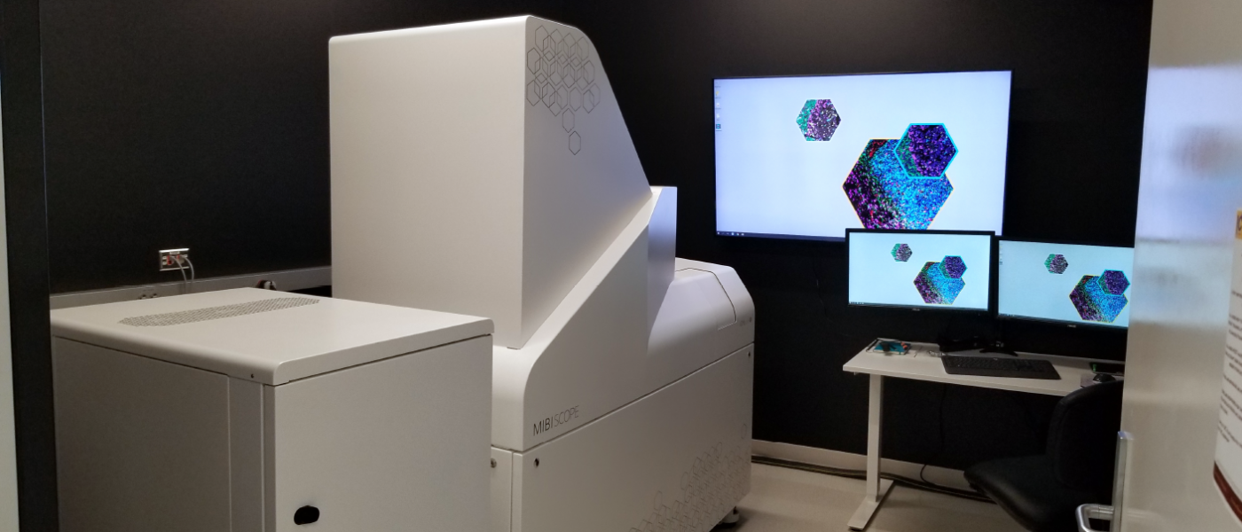
Multiplexed Ion-Beam Imaging (MIBI) is a powerful technique used to probe the microenvironment of pathological tissues at the single-cell level. With Ionpath’s MIBIscope, we can combine up to 40 markers to produce high-resolution images at the molecular level. Our unbiased analytical approach leverages the most recent advances in machine learning to provide reliable results and easy to interpret reports. Our analytical toolbox is continually evolving to better support the MIBIscope platform, building on these fundamentals:
- Quantify marker expression on a single-cell basis
- Create immune profiles and hierarchical trajectory inference
- Profile tissue architecture with spatial analysis
- Discover novel phenotypes
For more information on our Research Service, please refer to this detailed brochure.
We Can Help You
Research Questions That May Be Answered With MIBI:
- How does cell composition change with the disease context?
- Does the expression or co-expression of cell markers change with disease context?
- Does cell or structural morphology vary with disease context?
- Are there any interactions between specific cell types within tissue, and does this change with disease context?
- Do cells localize to histological structures and does this vary with disease context?
- What is the role of the cell microenvironment in a diseased setting?
What We Offer
What we offer is a Research Service that includes everything from experimental design and staining of specimens to reporting results using advanced computation methods.
- Panel selection and validation
- Image acquisition, processing and segmentation
- Unbiased machine learning tools for:
- Single-cell analysis
- Immune-profiling
- Spatial analysis
- Differential analysis
- Turn-key and custom analysis pipelines
- Easy to interpret reports
MIBIscope enables high-definition spatial proteomics, and is a powerful research tool for visualization, quantitation and discovery.
How We Do It
We have built an automated analysis pipeline for MIBI images that incorporates peer-reviewed algorithms that have arisen out of a need for handling high-dimensional datasets that would otherwise be next to impossible to perform manually. Spatial analysis of these images can identify proximity and relative spatial distribution of cells and features across a tissue to provide context that is otherwise not possible from traditional sequencing and cytometry datasets.
The pipeline is wholly customizable to the research question being asked. It is built on a framework of validated methods for sample preparation, data pre-processing and population identification. The objective is to both explain the experiment in an interpretable manner and to provide insight into the discovery of novel mechanisms.
Contact Us
Jamie McNicol, MIBI Data Scientist and Operator
Email: jmcnic@mcmaster.ca